 Loading... Please wait...
Popular Brands
Our Newsletter
 New Products
-
 $7,250.00
-
 $5,400.00
-
 $6,700.00
-
 $10,200.00
-
 $11,700.00
- Home
- Medical & Lab Equipment, Devices
- Philips HD11xe Ultrasound System, Includes S4-1 & L12-5 Probes
- Home
- Medical/Lab Equipment Attachments & Accessories
- Philips HD11xe Ultrasound System, Includes S4-1 & L12-5 Probes
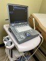
Philips HD11xe Ultrasound System, Includes S4-1 & L12-5 Probes

Product Description
Brand: Philips
MPN: 989605325131
Model: HD11 XE
Serial Number: us10771609
Philips HD11xe Ultrasound System
Philips Medical is known for bringing some of the best imaging systems to the market. They continue in this tradition of excellence with the Philips HD11 Imaging System. It's unique because it's more than just an Ultrasound. It can expand to a complete, digital cardiovascular system and has options including QLAB advanced, quantification, stress echo, contrast imaging and additionally, ultrasound guided regional anesthesia and emergency medicine configurations.
Additional features of this system include:
SonoCT – this feature creates exceptionally clear images because of the beam-steered spatial compounding. In an independent clinical study, it was determined that there was an increase in image superiority of 94% which led to changing patient treatment plans in some cases. (Scientifica study on image quality).
XRES – this feature dramatically lessens speckle and other types of noise artifact which clarifies each ultrasound image ensuring accurate diagnosis.
2D with Pulse Inversion Harmonic Imaging – as a patented method for producing the clearest images, the broadband harmonic signals enhance the image for ultimate viewing3D Imaging – the system offers "multiplanar views for qualitative 3D images" and interacts with at least three anatomical planesAdaptive.
Color Doppler – this automated feature allows the healthcare provider the ability to see Doppler or Angio frequency for images with sensitive resolution. This is technology for assessing amplitude, direction and flow.
Pulsed Wave and Continuous Wave Doppler – Adaptive Doppler technology that enhances signals that are typically weak and produce additional artifact.
Higher PRF to view and measure higher velocities than ordinary pulsed.
Doppler Contrast Imaging – Allows the Healthcare provider the ability to detect harmonic agents using the S3-1 and C5-2 transducers. This excites the image and contrast agents in the viewing region. This feature also reduces the need for system adjustments which make for a more complete visualization of contrast.
Panoramic Imaging – This provides an extended view of display. Creates real-time images while the transducer moves laterally across the anatomical region. Once imaging is completed, the system will automatically provide a panoramic mosaic image. This is unique as it provides a larger reference to aid in diagnosis.
Data Management and Connectivity – There are on-screen thumbnails to assist in building the patient study and overall exam status, at one glance. Additionally, there's a simple archiving feature that meets strict documentation requirements. This feature also allows you to quickly create patient reports with images embedded within the report.
DICOM Networking Option including Print and Store, Modality Worklist, Performed Procedure Step and Structured Reporting.